")
The phrase “life under the microscope” evokes a sense of wonder and revelation. It refers to the astonishing realm of tiny living things and structures that become visible only when magnified hundreds or thousands of times. This invisible world includes single-celled organisms, intricate cellular components, and even the molecular machinery that sustains life. What appears to the unaided eye as still water, a leaf, or a drop of blood transforms into bustling ecosystems of motion, interaction, and complexity. Exploring this domain has reshaped our understanding of biology, health, ecology, and even our place in the natural order. Here, we’ll examine the tools that open this world, the forms of life it reveals, their profound roles, the historical breakthroughs that brought them to light, and the ongoing frontiers that continue to expand our knowledge, all presented in an original synthesis drawn from core scientific concepts.
 The ability to see beyond normal vision depends on magnification and resolution. Early microscopes, developed in the 17th century, used ground lenses to bend light and enlarge images. Simple single-lens devices could reach 200–300× magnification, sufficient to observe bacteria for the first time. Compound microscopes, stacking multiple lenses, improve clarity and power. Modern optical microscopes use visible light and achieve up to about 1,500×, limited by the wavelength of light itself (around 0.2 micrometers resolution). To overcome this, electron microscopes fire streams of electrons, which have much shorter wavelengths, yielding resolutions down to fractions of a nanometer, enough to distinguish individual atoms in some cases.
Different types serve distinct purposes. Brightfield microscopes illuminate specimens from below for basic viewing of stained slides. Darkfield setups scatter light to highlight edges, making transparent organisms glow against a black background. Phase-contrast enhances subtle differences in refractive index, ideal for living, unstained cells. Fluorescence microscopy excites fluorescent tags attached to specific molecules, lighting up DNA, proteins, or organelles in vivid colors. Confocal variants use lasers and pinholes to create sharp, three-dimensional slices, eliminating out-of-focus blur. At the extreme end, scanning and transmission electron microscopes provide breathtaking surface details or internal ultrastructure, though they require prepared, non-living samples.
Through these instruments, we encounter prokaryotic life—bacteria and archaea. These ancient, simple cells lack a nucleus, with genetic material coiled in a central region. Their shapes vary: spherical cocci cluster like grapes, rod-shaped bacilli align in chains, spiral forms twist elegantly. Many possess whip-like flagella for propulsion or hair-like pili for surface attachment. Under high magnification, their rigid cell walls stand out, especially when stained to differentiate thick peptidoglycan layers from thinner, membrane-protected ones. These microbes inhabit virtually every niche: boiling hot springs, acidic mine drainage, frozen polar soils, and the depths of oceans. They recycle nutrients by breaking down dead matter, fixing atmospheric nitrogen into forms plants can use, and participating in symbiotic relationships that sustain larger organisms.
Eukaryotic cells present greater complexity. Animal cells feature a flexible outer membrane enclosing a prominent nucleus that safeguards DNA. Surrounding it, mitochondria, often called power plants, generate energy through intricate folds called cristae. The endoplasmic reticulum weaves through the cytoplasm, studded with ribosomes that assemble proteins. The Golgi apparatus modifies and packages these molecules for export. Lysosomes act as recycling centers, digesting waste. In plant cells, rigid walls of cellulose provide structure, while chloroplasts capture sunlight to convert carbon dioxide and water into sugars and oxygen. Large central vacuoles store water, nutrients, and pigments, maintaining turgor pressure. Fungal cells share eukaryotic traits but build walls from chitin, forming branching networks of hyphae that spread through substrates.
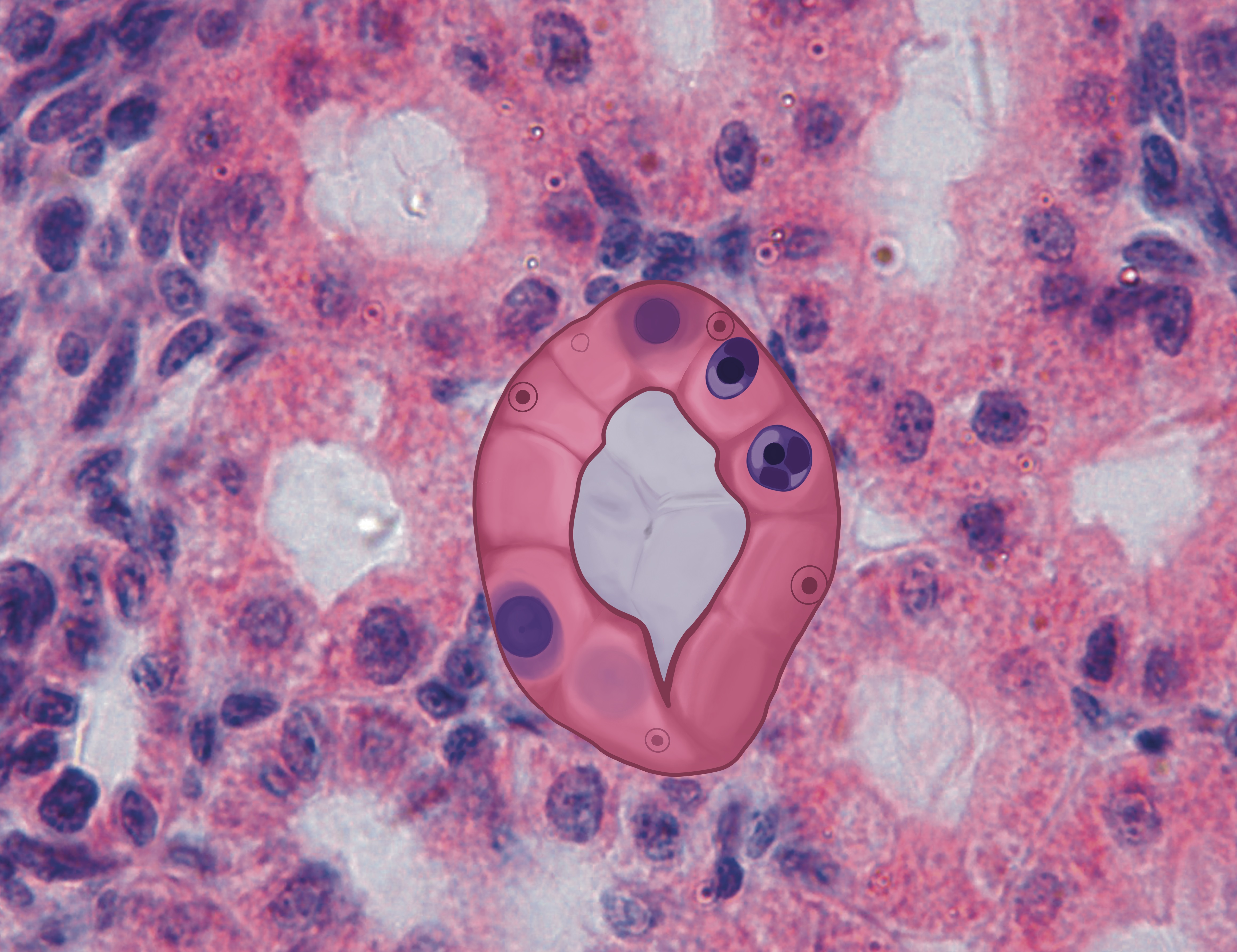
The Cell Through the Microscope – Anatomy and Physiology I: An Interactive Histology Atlas
Single-celled eukaryotes add diversity to the microscopic scene. Amoebas flow across slides using temporary extensions called pseudopods to engulf prey. Paramecia dart about, propelled by thousands of hair-like cilia, feeding through an oral groove. Ciliates and flagellates display coordinated movement, while some protists form colonies or intricate shells of silica or calcium carbonate. These organisms often appear in pond water samples, creating miniature dramas of predation, reproduction by binary fission or conjugation, and response to stimuli.
Viruses, though not cellular, dominate headlines and research. They consist of genetic material encased in protein coats, sometimes with lipid envelopes. Shapes range from simple icosahedral symmetry to complex tailed structures that infect bacteria. Under electron magnification, their geometric precision stands out, a reminder that life at its most basic level follows elegant patterns dictated by physics and chemistry.
SARS-CoV-2 under electron microscope | Colorado Encyclopedia
The impact of this hidden world extends far beyond observation. Microorganisms form the foundation of food webs: photosynthetic microbes in oceans produce a substantial portion of planetary oxygen and serve as base-level producers. In soils, bacterial and fungal communities enhance nutrient availability for plants while decomposing organic debris to recycle elements. The human body hosts a vast microbiome, trillions of microbes that aid digestion, produce essential vitamins, modulate immune responses, and even influence mood through gut-brain signaling. Disruptions to these communities can lead to health issues, from digestive disorders to weakened defenses against pathogens.
Historically, this realm revolutionized medicine. Observations of “little animals” in bodily fluids linked microbes to infection, paving the way for hygiene practices, sterilization, and targeted treatments. Antibiotics derived from microbial secretions combat bacterial diseases, while vaccines mimic natural immune responses to viruses. Diagnostic microscopy remains essential: examining blood for parasites, tissue for abnormal cell growth, or fluids for infectious agents.
Environmentally, microscopic life offers hope for sustainability. Certain bacteria degrade pollutants in contaminated water or soil, converting toxins into harmless byproducts. Algae and cyanobacteria hold promise for biofuel production, capturing carbon while generating energy-rich compounds. Engineered microbes synthesize pharmaceuticals, plastic alternatives, or enzymes for industrial processes.
Challenges persist. Antibiotic-resistant strains emerge from overuse, complicating treatments. Climate shifts alter microbial distributions, potentially unleashing dormant threats or changing disease patterns. Emerging pathogens remind us of vulnerability to unseen agents.
Advancements continue to push boundaries. Techniques that bypass light’s limits allow visualization of molecular dynamics in living cells. High-speed imaging captures processes in real time, revealing how cells divide, migrate, or respond to signals. Sequencing technologies uncover the genetic diversity of unculturable microbes, expanding our catalog of life’s forms. Artificial intelligence analyzes vast image datasets, identifying patterns humans might miss.
Ultimately, peering into this miniature universe fosters appreciation for life’s ingenuity. Every cell operates as a self-contained factory, coordinating thousands of reactions with precision. Communities of microbes collaborate in ways that mirror larger ecosystems. This interconnectedness underscores a fundamental truth: the smallest entities sustain the largest systems. By continuing to explore life under the microscope, we gain tools to heal, protect the environment, and deepen our understanding of existence itself. The view through the lens is not just magnification; it’s an invitation to humility and curiosity in the face of profound complexity.